ABSTRACT
Morphological changes occurring in Silk Fibroin -Rhodamine 6G films were examined using scanning electron microscopy (SEM). SEM analysis revealed that treatment of SF films
with 70% methanol induces the formation of large aggregates. However, in the presence of RHD 6G, these protein aggregates appear to be fragmented into significantly smaller
structures. X-ray diffraction (XRD) and Fourier-transform infrared (FTIR) spectroscopy further indicate that prolonged exposure of SF films to 70% methanol results in
dehydration, leading to the dense packing of SF amyloids.
Keywords: silk fibroin, amyloid, aggregation, neurodegenerative diseases, fluorescence, SEM, XRD
DOI:10.70784/azip.2.2025251
Received: 21.05.2025
Internet publishing: 17.06.2025 AJP Fizika A 2025 02 az p.51-55
AUTHORS & AFFILIATIONS
Institute of Biophysics, Ministry of Science and Education Republic of Azerbaijan 117 Z. Khalilov, Baku, Azerbaijan
E-mail: a.k134@hotmail.com
Graphics and Images
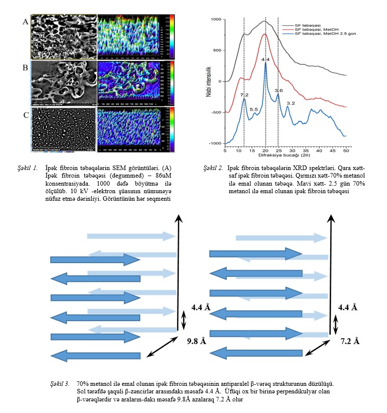
Fig.1-2-3
|
[1] F. J. O’Brien. “Biomaterials & scaffolds for tissue engineering,” Mater. Today, vol. 14, № 3, pp. 88–95, 2011. doi: 10.1016/S1369-7021(11)70058-X.
[2] C. Vepari and D.L. Kaplan. “Silk as biomaterial,” Prog. Polym. Sci., vol. 100, № 2, pp. 130–134, 2012, doi: 10.1016/j.progpolymsci.2007.05.013.Silk
[3] S. Kapoor and S.C. Kundu. “Silk protein-based hydrogels: Promising advanced materials for biomedical applications,” Acta Biomater., vol. 31, pp. 17–32, 2016, doi: 10.1016/j.actbio.2015.11.034.
[4] L. Jean, A.C. Foley and D.J.T. Vaux. “The physiological and pathological implications of the formation of hydrogels, with a specific focus on amyloid polypeptides,” Biomolecules, vol. 7, № 4, 2017, doi: 10.3390/biom7040070.
[5] O.K. Gasymov, C. Botta, L. Ragona, A.J. Guliyeva and H. Molinari. “Silk Fibroin-Based Films Enhance Rhodamine 6G Emission in the Solid State: A Chemical-Physical Analysis of their Interactions for the Design of Highly Emissive Biomaterials,” Macromol. Chem. Phys., vol. 220, № 4, 2019, doi: 10.1002/macp.201970007.
[6] V. Murugesan, S. Mundada and S. Rajagopal. “Silk fibroin as a functional biomaterial for tissue engineering,” Silk Fibroin Adv. Appl. Res., pp. 155–175, 2023.
[7] A. Ogunsipe. “Solvent Effects on the Spectral Properties of Rhodamine 6G: Estimation of Ground and Excited State Dipole Moments,” J. Solution Chem., vol. 47, № 2, pp. 203–219, 2018, doi: 10.1007/s10953-017-0706-8.
[8] S. Tomaselli et al. “Encapsulation of a rhodamine dye within a bile acid binding protein: Toward water processable functional bio host-guest materials,” Biomacromolecules, vol. 14, № 10, pp. 3549–3556, 2013, doi: 10.1021/bm400904s.
[9] R.F. Həşİmov. La0.63Ba0.37MnO3 birləşməsinin kristal quruluşununv rentgenoqrafik tədqiqi. AJP Fizika, cild XXVIII, № 1, section: Az, pp. 35–37, 2022.
[10] A.M. Mammedzade, B.U. Gasimli. Structural origin of silk nanoparticles and their stabilization. AJP Fizika, vol. XXIX, section En, № 3, pp. 6–12, 2023.
[11] W. Huang, S. Ling, C. Li, F.G. Omenetto, and D.L. Kaplan. “Silk-based Materials and Devices through Bio-nanotechnology Article Type: Tutorial Review Chemical Society Reviews Article type: Tutorial Review Paper Title: Silkworm Silk-based Materials and Devices generated using Bio-nanotechnology,” 2018.
[12] B.D. Lawrence et al.. “Effect of hydration on silk film material properties,” Macromol. Biosci., vol. 10, № 4, pp. 393–403, 2010, doi: 10.1002/mabi.200900294.
[13] B.D. Lawrence, M. Cronin-Golomb, I. Georgakoudi, D.L. Kaplan, and F.G. Omenetto. “Bioactive silk protein biomaterial systems for optical devices,” Biomacromolecules, vol. 9, № 4, pp. 1214–1220, 2008, doi: 10.1021/bm701235f
[14] H.J. Jin et al.. “Water-stable silk films with reduced β-sheet content,” Adv. Funct. Mater., vol. 15, № 8, pp. 1241–1247, 2005, doi: 10.1002/adfm.200400405.
[15] A.S. Cohen and M. Skinner. “the Amyloid Fibril,” № 34110, pp. 1373–1375.
[16] L.C. Serpell, J. Berriman, R. Jakes, M. Goedert, and R.A. Crowther. “Fiber diffraction of synthetic α-synuclein filaments shows amyloid-like cross-β conformation,” Proc. Natl. Acad. Sci. U. S. A., vol. 97, № 9, pp. 4897–4902, 2000, doi: 10.1073/pnas.97.9.4897.
[17] K.L. Morris and L.C. Serpell. “X-ray fibre diffraction studies of amyloid fibrils,” Methods Mol. Biol., vol. 849, pp. 121–135, 2012, doi: 10.1007/978-1-61779-551-0_9.
[18] S. Dutta, B. Talukdar, R. Bharali, R. Rajkhowa, and D. Devi. “Fabrication and characterization of biomaterial film from gland silk of muga and eri silkworms,” Biopolymers, vol. 99, № 5, pp. 326–333, 2013, doi: 10.1002/bip.22168.
[19] G.G. Kariyappa et al.. “Crystallite Shapes and Functional Data Analysis of Silk forms using X-ray Diffraction: Microwave Irradiation Effects,” Biointerface Res. Appl. Chem., vol. 13, № 3, pp. 1–10, 2023, doi: 10.33263/BRIAC133.241.
|